Welcome to the Radius and Ulna Labeling Quiz! This interactive challenge will put your understanding of forearm anatomy to the test. Whether you’re a medical student, an anatomy enthusiast, or simply curious about the human body, this quiz is designed to engage and educate you.
As you embark on this journey, you’ll delve into the fascinating world of the radius and ulna bones, exploring their unique features, anatomical relationships, and clinical significance. So, grab a pen and paper, or simply get ready to flex your mental muscles, and let’s begin!
Overview of Radius and Ulna Bones
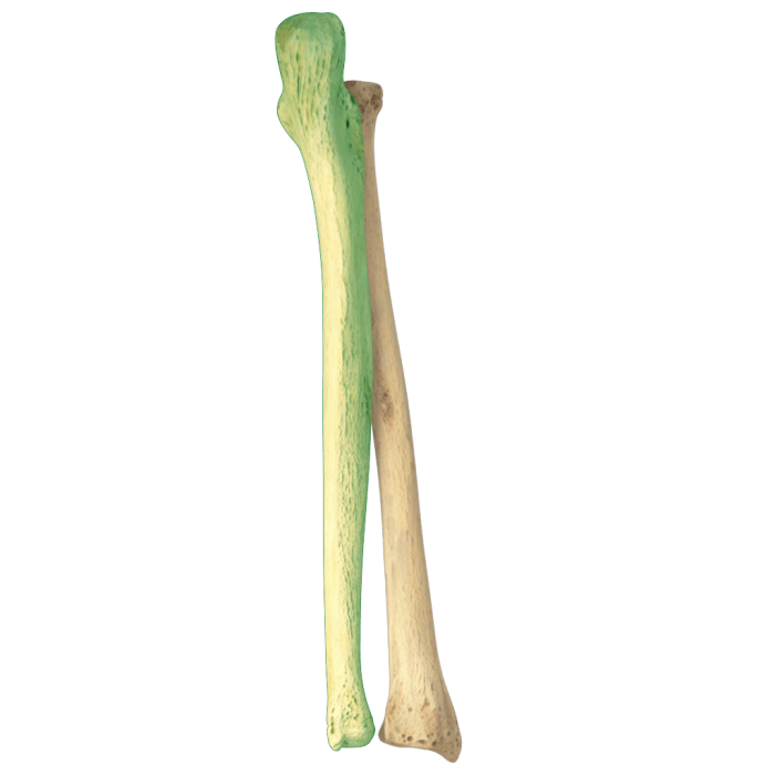
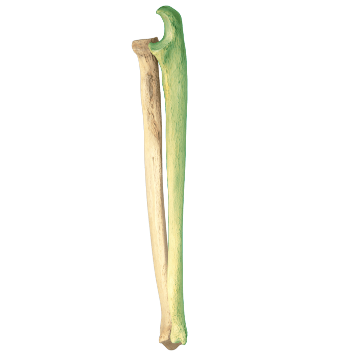
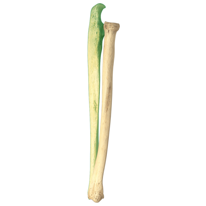
The radius and ulna are two long bones that form the forearm. They are located on the lateral (thumb side) and medial (little finger side) sides of the forearm, respectively.
Ready for the radius and ulna labeling quiz? To prepare, check out the unit 1 progress check: frq . The practice questions will help you master the concepts and ace the quiz on radius and ulna labeling.
The radius is the shorter and thicker of the two bones. It articulates with the humerus (upper arm bone) at the proximal end and with the carpal bones (wrist bones) at the distal end. The ulna is the longer and thinner of the two bones.
It articulates with the humerus at the proximal end and with the radius at the distal end.
The radius and ulna work together to allow for a wide range of motion in the forearm, including pronation (turning the palm down) and supination (turning the palm up).
Anatomical Position
In anatomical position, the radius and ulna are held parallel to each other, with the radius on the lateral side and the ulna on the medial side. The proximal ends of the radius and ulna articulate with the humerus at the elbow joint.
The distal ends of the radius and ulna articulate with the carpal bones at the wrist joint.
Relationship to Other Bones
The radius and ulna articulate with the following bones:
- Humerus (upper arm bone)
- Carpal bones (wrist bones)
Distinguishing Features of Radius and Ulna
The radius and ulna are two parallel bones in the forearm that are connected by the interosseous membrane. Despite their close proximity, they have distinct features that differentiate them from each other.
The radius is the lateral bone of the forearm, while the ulna is the medial bone. The radius is typically longer and thicker than the ulna, and it has a more curved shape. The ulna, on the other hand, is flatter and straighter.
Shape and Size, Radius and ulna labeling quiz
The radius is a long bone with a slightly curved shaft. The ulna is also a long bone, but it is flatter and straighter than the radius. The radius is typically longer than the ulna, and it is thicker at its proximal end.
Surface Anatomy
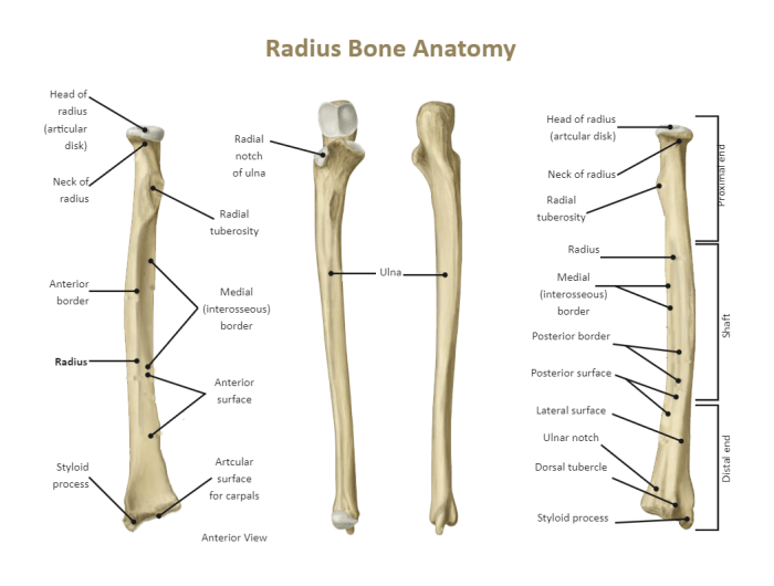
The radius has a smooth, rounded surface, while the ulna has a rougher, more irregular surface. The radius has a prominent ridge called the radial tuberosity, which is located near the proximal end of the bone. The ulna has a similar ridge called the olecranon process, which is located at the proximal end of the bone.
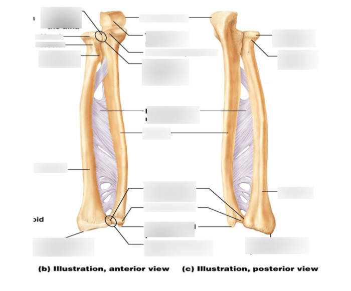
Labeling the Radius and Ulna: Radius And Ulna Labeling Quiz
Test your knowledge of radius and ulna anatomy with our interactive labeling quiz. Simply click on the correct label for each structure, and we’ll provide instant feedback.
This quiz is a great way to reinforce your understanding of the radius and ulna, and to prepare for exams or clinical practice.
Interactive Labeling Quiz
- Drag the label “Radius” to the correct location on the image.
- Drag the label “Ulna” to the correct location on the image.
- Drag the label “Radial head” to the correct location on the image.
- Drag the label “Radial neck” to the correct location on the image.
- Drag the label “Radial tuberosity” to the correct location on the image.
- Drag the label “Ulna styloid process” to the correct location on the image.
- Drag the label “Trochlear notch” to the correct location on the image.
- Drag the label “Coronoid process” to the correct location on the image.
- Drag the label “Olecranon process” to the correct location on the image.
Clinical Significance of Radius and Ulna Injuries
Injuries to the radius and ulna, collectively known as forearm fractures, are prevalent and can significantly affect an individual’s mobility and daily functioning. These injuries can range from simple fractures to complex, displaced breaks.
Common Radius and Ulna Injuries
- Distal Radius Fractures:These are the most common type of forearm fractures, often occurring due to falls or direct impact on the wrist. They can involve the distal end of the radius bone, including the styloid process.
- Radial Shaft Fractures:Fractures along the shaft of the radius bone are usually caused by direct trauma or a fall on an outstretched arm. They can be isolated or associated with other forearm injuries.
- Ulna Shaft Fractures:Isolated fractures of the ulna shaft are less common than radius fractures. They often result from a direct blow to the forearm or a fall on the hand.
- Monteggia Fracture:This complex injury involves a fracture of the ulna shaft with a dislocation of the radial head at the elbow joint. It can be caused by a fall or a forceful blow to the forearm.
- Galeazzi Fracture:Another complex injury, it involves a fracture of the radius shaft with a dislocation of the distal ulna at the wrist joint. It is typically caused by a fall on an outstretched hand.
Impact of Radius and Ulna Injuries on Function and Movement
Forearm fractures can significantly impair an individual’s ability to perform daily tasks and engage in physical activities. These injuries can cause:
- Pain and Swelling:Fractures result in pain, inflammation, and swelling in the forearm, which can limit mobility.
- Loss of Range of Motion:The pain and swelling can restrict the range of motion in the wrist, elbow, and shoulder joints.
- Grip Strength Weakness:Fractures in the forearm can weaken the grip strength, making it difficult to hold objects or perform activities that require hand strength.
- Nerve Damage:In severe cases, fractures can damage the nerves in the forearm, leading to numbness, tingling, or weakness in the hand and fingers.
Additional Resources for Learning
Enhance your understanding of radius and ulna anatomy with these valuable resources:
Websites:
- Kenhub:Interactive 3D models and detailed explanations of radius and ulna anatomy.
- TeachMeAnatomy:Comprehensive diagrams and videos showcasing the structures and functions of radius and ulna.
- Anatomy & Physiology Course:Free online course from Lumen Learning covering radius and ulna anatomy, including quizzes and interactive simulations.
Textbooks:
- Gray’s Anatomy for Students:The classic reference for human anatomy, providing detailed descriptions and illustrations of radius and ulna.
- Moore’s Clinically Oriented Anatomy:Focuses on the clinical relevance of radius and ulna anatomy, including common injuries and treatments.
- Grant’s Atlas of Anatomy:A comprehensive atlas with high-quality images and illustrations of radius and ulna anatomy.
Videos:
- Radius and Ulna Anatomy:Crash Course video providing a concise overview of radius and ulna anatomy and their functions.
- Radius and Ulna Bone Tutorial:MedSchool Insiders video explaining the anatomy and movements of radius and ulna.
- Radius and Ulna Fracture Management:Orthopaedic Surgery Essentials video discussing common fractures of radius and ulna and their management.
Questions Often Asked
What is the radius bone?
The radius is the lateral bone of the forearm, located on the thumb side.
What is the ulna bone?
The ulna is the medial bone of the forearm, located on the little finger side.
What is the function of the radius and ulna bones?
The radius and ulna bones work together to provide stability, support, and movement to the forearm.
What are common injuries associated with the radius and ulna?
Common injuries include fractures, sprains, and dislocations.